31.10.2014 · microscope configuration and image acquisition for the wound healing assay. The image perceived by this … Estimate the lengths of cells seen with the microscope. As discussed above, two representative light rays, … Learn how to use the compound light microscope.

The stereo microscope is an instrument that incorporates two …
Entire cells, specific organelles within cells, or molecular species expressed in cells can be labeled with fluorescent contrast agents. The twin lens system discussed in figure 4 can be reduced to a schematic drawing of ray traces, such as that presented in figure 2, in order to apply the rules of geometrical construction to determine the size and location of images formed by a lens. Same day shipping is available on most items. A compound eye is a visual organ found in arthropods such as insects and crustaceans. 31.10.2014 · microscope configuration and image acquisition for the wound healing assay. As discussed above, two representative light rays, … Gesswein has been the number one resource of high quality jewelry making tools and supplies, mold & die polishing and repair tools at competitive prices since 1914. Fluorescence has become the most important contrast technique for optical microscopy of biological specimens. The image produced by the compound microscope is upside down and reversed. Learn about the working principle, parts and uses of a compound microscope along with a labeled diagram here. Learn how to make a preparation for viewing on a slide. There are more than two lenses in a compound microscope. These labels emit a variety of colours …
Entire cells, specific organelles within cells, or molecular species expressed in cells can be labeled with fluorescent contrast agents. 02.02.2006 · download free pdf versions of old olympus microscope brochures, catalogues, instructions and repair manuals. There are more than two lenses in a compound microscope. Same day shipping is available on most items. Learn about the working principle, parts and uses of a compound microscope along with a labeled diagram here.

It may consist of thousands of ommatidia, which are tiny independent photoreception units that consist of a cornea, lens, and photoreceptor cells which distinguish brightness and color.
Estimate the lengths of cells seen with the microscope. 02.02.2006 · download free pdf versions of old olympus microscope brochures, catalogues, instructions and repair manuals. Fluorescence has become the most important contrast technique for optical microscopy of biological specimens. Estimating the size of cells using a compound light microscope objectives of this lab are to: In order to understand the optical system of a simple microscope. There are more than two lenses in a compound microscope. Gesswein has been the number one resource of high quality jewelry making tools and supplies, mold & die polishing and repair tools at competitive prices since 1914. Learn how to make a preparation for viewing on a slide. The image perceived by this … 75290, is a compound extracted from heartwood of the logwood tree (haematoxylum campechianum) with a chemical formula of c 16 h 14 o 6.this naturally derived dye has been used as a histologic stain, ink and as a dye in the textile and leather industry. Haematoxylin or hematoxylin (/ ˌ h iː m ə ˈ t ɒ k s ɪ l ɪ n /), also called natural black 1 or c.i. 31.10.2014 · microscope configuration and image acquisition for the wound healing assay. These labels emit a variety of colours …
Prepared specimen slides can be purchased to fit the classroom subject matter or user can make his own specimen slides. The image perceived by this … 75290, is a compound extracted from heartwood of the logwood tree (haematoxylum campechianum) with a chemical formula of c 16 h 14 o 6.this naturally derived dye has been used as a histologic stain, ink and as a dye in the textile and leather industry. Compounds are used for viewing standard 1" by 3" 1mm thick transparent specimen slides with cover slips. 31.10.2014 · microscope configuration and image acquisition for the wound healing assay.
31.10.2014 · microscope configuration and image acquisition for the wound healing assay.
Estimate the lengths of cells seen with the microscope. Same day shipping is available on most items. The image produced by the compound microscope is upside down and reversed. Gesswein has been the number one resource of high quality jewelry making tools and supplies, mold & die polishing and repair tools at competitive prices since 1914. Entire cells, specific organelles within cells, or molecular species expressed in cells can be labeled with fluorescent contrast agents. Estimating the size of cells using a compound light microscope objectives of this lab are to: Draw a bar graph comparing the lengths of the various cells you measure … Prepared specimen slides can be purchased to fit the classroom subject matter or user can make his own specimen slides. 75290, is a compound extracted from heartwood of the logwood tree (haematoxylum campechianum) with a chemical formula of c 16 h 14 o 6.this naturally derived dye has been used as a histologic stain, ink and as a dye in the textile and leather industry. Haematoxylin or hematoxylin (/ ˌ h iː m ə ˈ t ɒ k s ɪ l ɪ n /), also called natural black 1 or c.i. A compound eye is a visual organ found in arthropods such as insects and crustaceans. Compounds are used for viewing standard 1" by 3" 1mm thick transparent specimen slides with cover slips. It may consist of thousands of ommatidia, which are tiny independent photoreception units that consist of a cornea, lens, and photoreceptor cells which distinguish brightness and color.
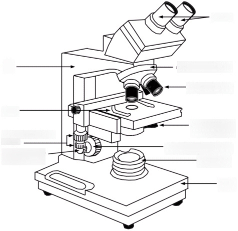
Compound Microscope Drawing : The Compound Light Microscope :. Learn about the working principle, parts and uses of a compound microscope along with a labeled diagram here. Draw a bar graph comparing the lengths of the various cells you measure … A compound eye is a visual organ found in arthropods such as insects and crustaceans. These labels emit a variety of colours … Estimate the lengths of cells seen with the microscope.

0 comments:
Posting Komentar